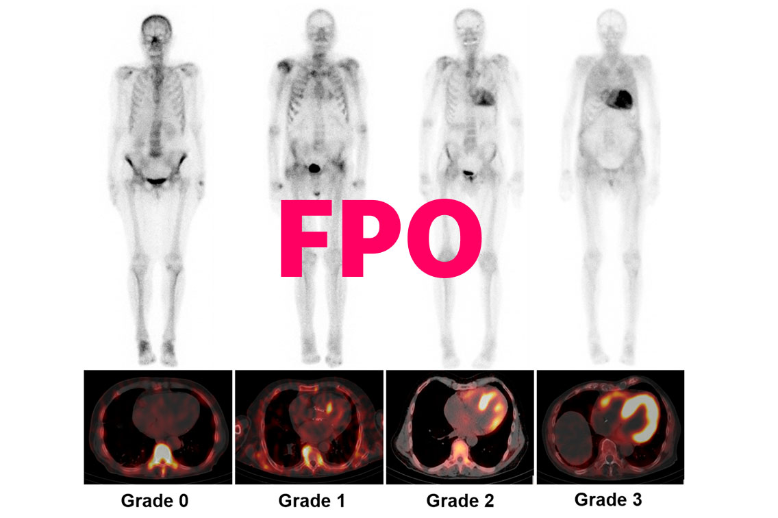
In this video, Dr Garcia-Pavia discusses the role of scintigraphy in diagnosing ATTR-CM, as well as how to interpret other diagnostic screening tests for ATTR-CM.
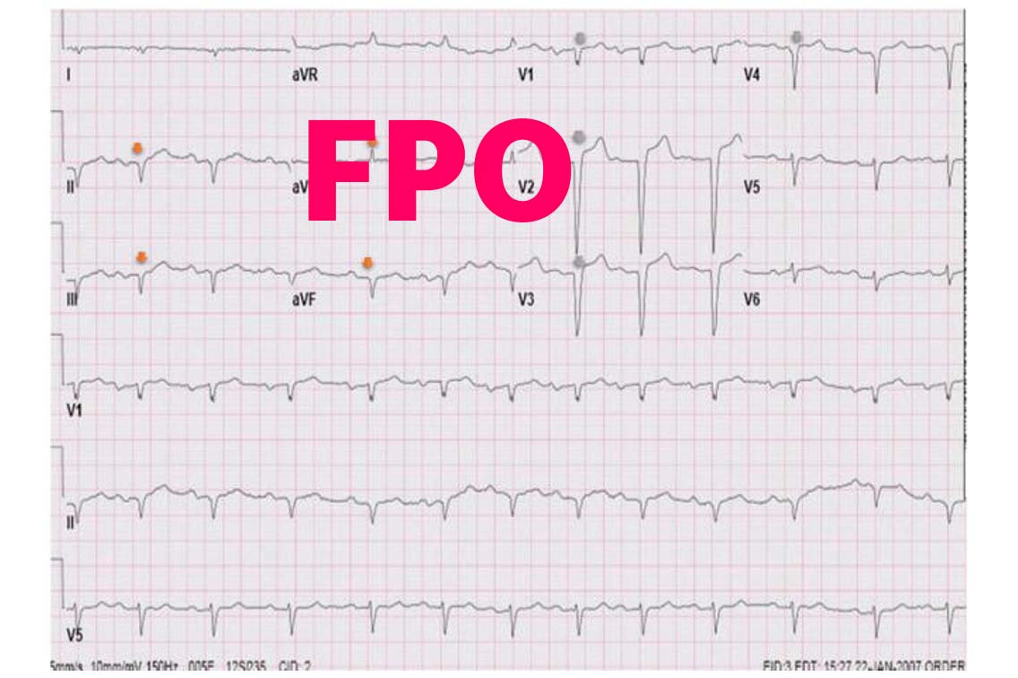
ECG4
- Pseudo-infarction pattern
- QRS voltages disproportionate to LV wall thickness
- Atrioventricular block in the presence of LVH
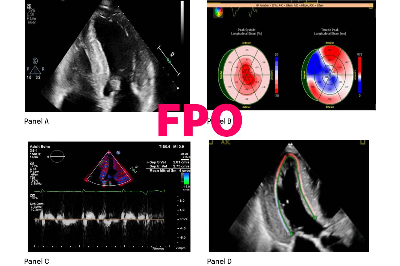
ECHO4
- Hypertrophic phenotype with infiltrative features
- Restrictive LV filling with RV wall thickening
- Low septal and lateral e′
- Reduction in global longitudinal strain
Proactively looking for these findings in regular clinical practice may facilitate an ATTR-CM diagnosis5
Once ATTR-CM is suspected, ACT FAST. Learn about the diagnostic process to confirm ATTR-CM


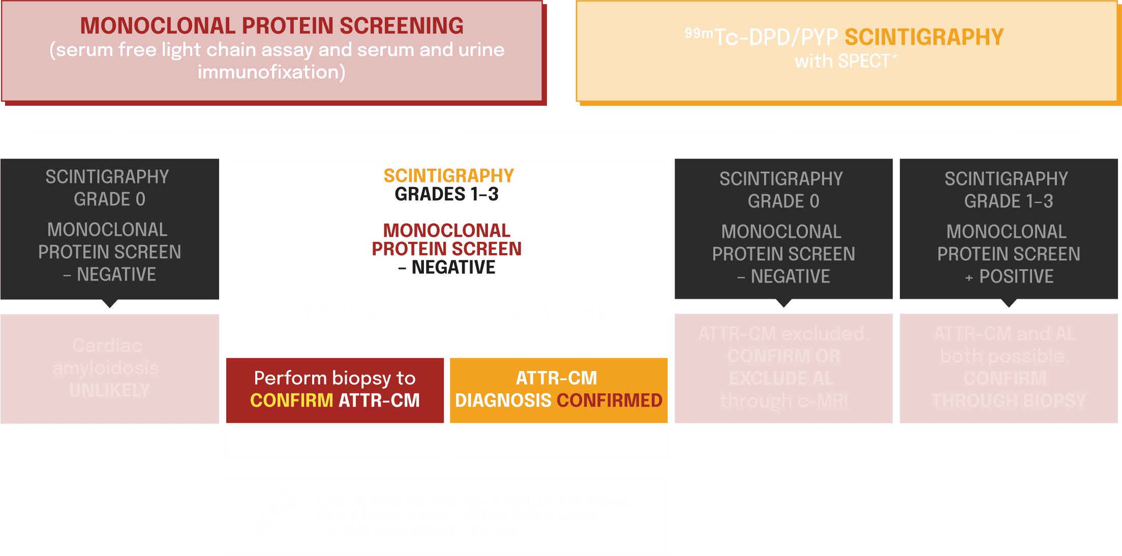
*99mTc-HMDP scintigraphy can also be used.6
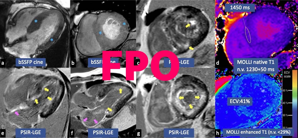
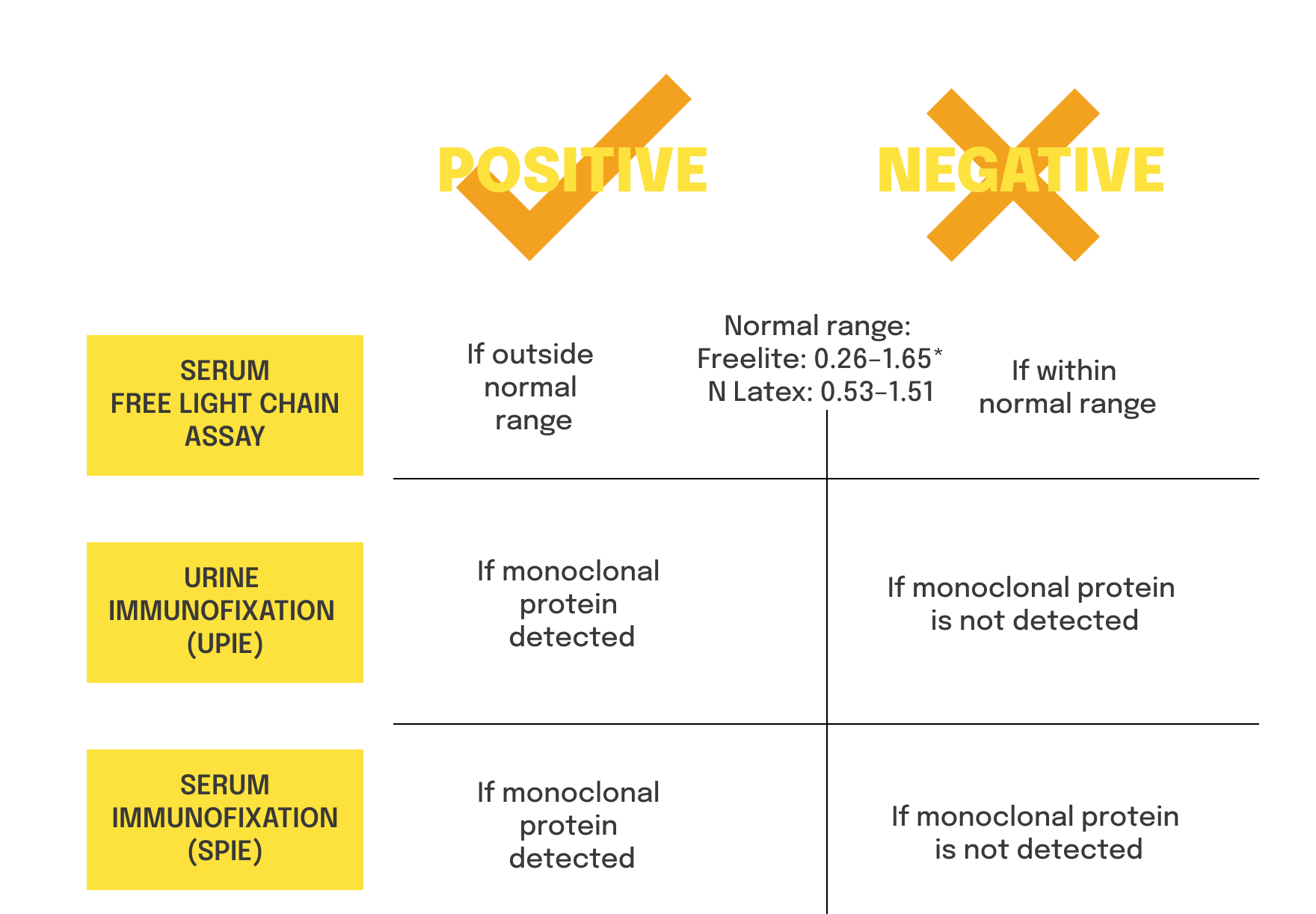
†Histological confirmation of amyloid deposits (can be cardiac or extracardiac) is required. If the biopsy is extracardiac, and accompanied with characteristic features on ECHO or c-MRI, diagnosis can be confirmed. Cardiac amyloidosis on biopsy is characterised by extracellular deposition of misfolded proteins with pathognomonic histological property of green birefringence when viewed under cross-polarised light after staining with Congo red.6
hATTR and wtATTR can present differently, and may require a
different treatment and management strategy1,6
Detection of a pathogenic TTR variant allows for
testing of family members
and earlier detection of symptoms and treatment intervention7,8
TREAT
Specific ATTR-CM treatments are available.9 Initiating these treatments as early as possible can lead to improved outcomes9
[Local information for guidance on treating ATTR-CM]
OR REFER
ATTR-CM is a rapidly progressive disease.4 It is vital to ACT FAST. Referral to a specialist centre may ensure patients get the right treatment, sooner
[Local information for guidance on treating ATTR-CM]
Once ATTR-CM is confirmed, ACT FAST. Learn about how patients may present in your clinic